
Introduction
Gnomonia leaf blotch occurs sporadically in annual strawberry production systems and is often associated with plant source. The pathogen is most commonly found on foliage in our region, on rare occasions it may infect flower parts and can cause stem end fruit lesions. It can build up in plug production facilities causing leaf blotches detracting from the look of the plug plants. However, the pathogen rarely causes economic damage.
Symptoms and Signs
Gnomonia infects leaves causing brownish to purplish lesions that begin small but expand to large areas, especially as lesions coalesce to form large blotchy areas of damage on leaves (Figure SS-1). Lesions can have various shades and are visible on the upper and lower side of the leaf (Figure SS-2). These spots often occur on the end of a leaflet and are V-shaped. As the disease progresses, the outer leaves of affected strawberry plants often die. Frequently, the lesions have small raised bumps, or pycnidia, visible with a 20 to 30x hand lens. If these bumps are not visible in the field, they emerge after a short time in incubation chambers (Figure SS-3). The pycnidia have a yellow to brown color, ostioles (holes on top) and exude small conidia that have two oil bodies, one at each end of the conidia (Figure SS-3). The pathogen can also form microscopic sexual structures visible as black long necked and flasked shaped structures called perithecia, often visible on leaf spots and petiole lesions.
Under favorable weather conditions, the pathogen causes a flower blast where flowers are heavily colonized by the pathogen turning the calyx, peduncle and other flower parts brown. In some cases, the pathogen colonizes the stem of the fruit causing stem-end rot (Figure SS-4) characterized by circular to irregularly shaped brown lesions. Fruit may be infected at all stages of development.
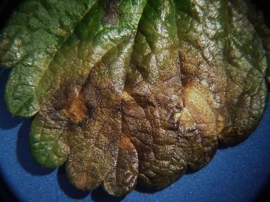
Figure SS-1: Gnomonia leaf blotch on strawberry leaf. Several lesions may coalesce to cause large blotches on leaves. Note small raised bumps (pycnidia) surrounding the lesions.
Frank J. Louws
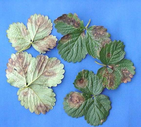
Figure SS-2: Gnomonia leaf blotch showing a range of symptoms on the lower and upper surface of the leaves.
Frank J. Louws
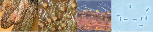
Figure SS-3: Gnomonia leaf blotch on leaf showing pycnidia around the leaf lesion appearing as raised bumps (~10x); pycnidia erupting from stem/peduncle lesions (middle ~50 x and 20x) and small conidia (5-6 * 2 ?m) with 2 oil bodies that refract light.
Frank J. Louws
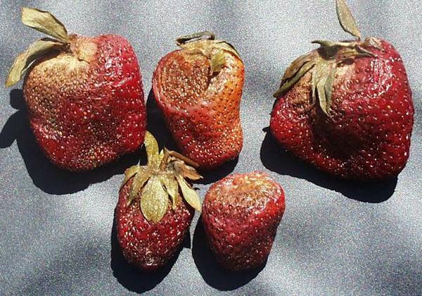
Figure SS-4: Gnomonia stem-end rot showing the brown circular to irregular lesions that form at the stem end of the fruit.
Frank J. Louws
Disease Cycle
The biology and ecology of the pathogen in North Carolina and surrounding area is not well documented. G. comari sensu lato affects numerous herbaceous Rosaceae genera including Fragaria, Comarum, Geum, Potentilla, Alchemilla, Agrimonia, Sanguisorba and also Epilobium hirsutum in the evening primrose family. However, in the majority of cases, the initial inoculum is introduced into plug facilities and fruiting fields with transplants. Lesions on these plants produce pycnidia that ooze conidia that are then splash dispersed. The conidia germinate and infect the plant through stomata or wounds.
Management
In most cases specific management recommendations are not needed since the disease rarely develops to a point where economic losses occur. However, lesions on leaves, the blotches, can develop to concerning levels on plugs and soon after transplanting in fruiting fields. These leaves may affect early plant establishment but new leaves develop quickly and the disease does not persist into the early spring or fruiting period. In perhaps one year of 20, persistent cool wet weather during flowering and early fruit development led to flower blast and fruit stem-end rot that impacted yield in some fields. Little data is available for this disease but products effective against Phomopsis leaf blight appear effective against this disease. Products, rates and timing are highlighted in the Strawberry IPM Guide.
Pathogen
Gnomonia comari is a fungal plant pathogen that causes a minor disease of strawberry. To reproduce, G. comari forms globose and beaked perithecia. The base of the perithecium (250-600 µm in diameter) is buried in the host tissue and the cylindrical neck (200-1200 µm long) protrudes from it. Many asci (20-35 x 3.5-8 µm) are produced within the perithecium and each ascus contains 8 ascospores. Mature ascospores (6.5-13 x 1.5-2.5 µm) are hyaline, straight or slightly curved and submedially septate. Ascospores of G. comari also contain conspicuous oil droplets and lack appendages (CABI, 2008; Maas, 1998).
The anamorph of G. comari, Zythia fragariae produces pycnidia that are yellowish brown, soft walled, and ostiolate with no conspicuous neck. The conidia (5-6 x 2 µm) are hyaline, contain two oil droplets, have rounded ends, and are borne on short unbranched conidiophores (Maas, 1998).
Diagnostic Procedures
Symptoms of leaf blotch could be confused with those of Verticillium wilt, as both diseases affect outer leaves of the strawberry plant. Foliar symptoms of G. comari also may be similar to the light brown necrotic spots caused by Phomopsis obscurans. While G. comari can be distinguished from Verticillium species by the presence of beaked perithecia or pycnidia in leaf lesions, and spores with discrete oil droplets, differentiating between Gnomonia and Phomopsis is more difficult. The fruiting structures and spores of G. comari and P. obscurans can look similar although Gnomonia pycnidia tend to be yellow to brown and Phomopsis pycnidia tend toward black; Phomopsis conidia do not have the prominent oil bodies. It is important to note that G. comari produces both perithecia, bearing asci with ascospores, and pycnidia from which conidia are borne while P. obscurans is an asexual fungus and can only produce pycnidia.
When signs of the pathogen are absent, incubating symptomatic leaves or leaflets for 24 to 48 hours in a moist chamber usually results in abundant sporulation of the fungus.
References
CABI. 2008. Gnomonia comari. Crop Protection Compendium. CAB International, Wallingford, UK.
Maas, J. L. 1998. Compendium of Strawberry Diseases, 2nd edition. APS Press. St. Paul, MN.
Publication date: May 14, 2014
Revised: Aug. 19, 2019
There is an alternate Spanish language version of this document here: Mancha de la hoja de Gnomonia y podredumbre del extremo del tallo de la fresa
N.C. Cooperative Extension prohibits discrimination and harassment regardless of age, color, disability, family and marital status, gender identity, national origin, political beliefs, race, religion, sex (including pregnancy), sexual orientation and veteran status.