
From the Field - Agronomy Notes
In this Brassica carinata (Ethiopian mustard) research update, we highlight the symptoms of calcium deficiency. These images are part of a project by the Southeast Partnership for Advanced Renewables from Carinata (SPARC) to develop a diagnostic series for the identification of nutrient disorders of Carinata. Carinata is an exciting new crop in the Southeast used for a wide variety of primary and secondary agricultural products including cover crops, feedstock, high protein meal, and jet fuel. It is similar in management to canola given both canola and carinata are winter annual Brassica oilseed crops. However, carinata oil is not edible.
Symptoms
Calcium (Ca) is bound as cellular structures in plants, and is consequently plant-immobile. This means symptomology will manifest on the new growth rather than older foliage. The first sign of calcium deficiency will be evident on the growing tip, showing a curling of the margin of the new leaves (Figure 1). These new, expanding leaves will appear to develop normally except for the leaf margin, which will curl either inward or upward resulting in a cupping of the leaf (Figure 2). The leaves may also have small brown spots randomly distributed toward the petiole near the margin of the leaf (Figure 3).
The next stage of calcium deficiency results in general interveinal chlorosis of the new growth, particularly near the petiole (Figure 4). As the developing leaves continue to expand, the center of the leaf will expand while the margin does not, resulting in a more dramatic cupping of the leaf and occasionally splitting or cracking near the leaf margin. In more advanced stages, the leaf will turn from interveinal yellowing to interveinal reddening or purpling (Figure 5).
The final and most advanced stages of calcium deficiency result in severely distorted growing tips which appear as spiked petioles (Figure 6). The growing tip will eventually die, resulting in the proliferation of axillary shoots (Figure 6). These shoots will also appear as unexpanded leaves or spiked stems. To ensure proper diagnosis the above material should be used in conjunction with a leaf tissue sample and/or field test.
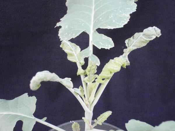
Figure 1. Note new, expanding leaves. They are exhibiting deformation especially along the margins. As calcium becomes limited, the leaf surface will continue to grow and expand; however, the leaf margin will remain stunted causing an inward curling.
Forensic Floriculture, 2018
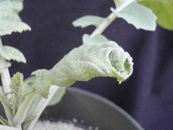
Figure 2. As calcium deficiency symptoms progress, the new leaves will continue to expand in the center however the leaf margin will remain undeveloped. This causes the leaf to distort and curl inward on itself forming a hood like structure.
Forensic Floriculture, 2018
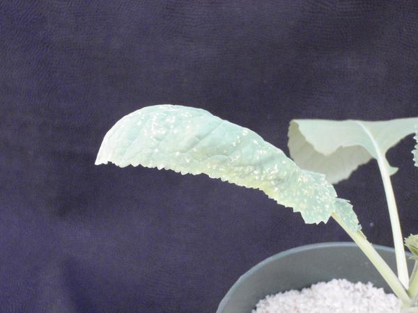
Figure 3. In addition to the marginal curling of the new foliage, the leaves will also develop necrotic brown to tan spots. These will become more severe and larger as symptoms progress.
Forensic Floriculture, 2018
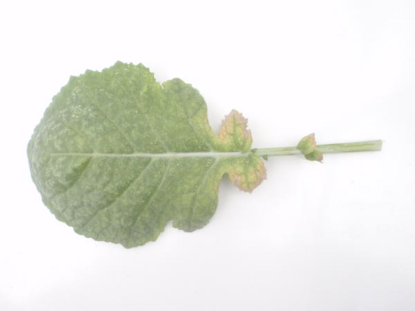
Figure 4. As calcium deficiency progresses, another indicator manifests as interveinal chlorosis especially near the petiole and leaf margin. This interveinal chlorosis will be accompanied with leaf distortion and small necrotic lesions.
Forensic Floriculture, 2018
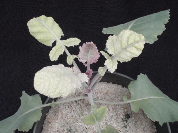
Figure 5. The expanding leaf above is displaying symptoms of marginal curling. In addition to these symptoms, note the red/purple/pink coloration of the newest foliage. This is another indicator of calcium deficiency conditions.
Forensic Floriculture, 2018
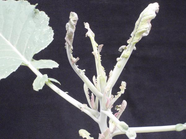
Figure 6. Eventually the calcium starvation will result in the death of the growing tip. This will cause the plant to start growth from the axillary shoots. As seen above, the axillary shoots are growing however they are severely distorted.
Forensic Floriculture, 2018
Project Support
We would like to thank the following for financial support of this project:


Key Contact
Key Contact Central East:
Dr. Angela Post, NC State Univ. Department of Crop and Soil Sciences – angela_post@ncsu.edu
Dr. Carl Crozier, NC State Univ. Department of Crop and Soil Sciences – ccrozier@ncsu.edu
Key Contact South East:
Dr. Michael Mulvaney, UF/IFAS West Florida Research and Education Center – m.mulvaney@ufl.edu
Primary Authors: Paul Cockson, Dr. Carl Crozier, Dr. Ramon Leon, Dr. Michael Mulvaney, Dr. Angela Post, and Dr. Brian E. Whipker
Project Team: NC State Univ. personnel Paul Cockson (NC State B.S. student in Agroecology), Ingram McCall (Research Technician in Horticultural Science at NC State), Dr. Carl Crozier (Professor and Extension Specialist at NC State), Dr. Ramon Leon (Assistant Professor at NC State), Dr. Angela Post (Assistant Professor and Extension Specialist NC State), and Dr. Brian Whipker (Professor of Floriculture and Plant Nutrition in Horticultural Science at NC State). Univ. of Florida personnel Dr. Michael Mulvaney (Cropping Systems Specialist at UF/IFAS West Florida Research and Education Center.
Publication date: Jan. 1, 2021
N.C. Cooperative Extension prohibits discrimination and harassment regardless of age, color, disability, family and marital status, gender identity, national origin, political beliefs, race, religion, sex (including pregnancy), sexual orientation and veteran status.